39 human cell diagram with labels
Anatomy and Physiology: Parts of a Human Cell - Visible Body The nucleus is a large organelle that contains the cell's genetic information. Most cells have only one nucleus, but some have more than one, and others—like mature red blood cells—don't have one at all. Within the nucleus is a spherical body known as the nucleolus, which contains clusters of protein, DNA, and RNA. Skeletal System - Labeled Diagrams of the Human Skeleton The skeletal system's cell matrix acts as our calcium bank by storing and releasing calcium ions into the blood as needed. Proper levels of calcium ions in the blood are essential to the proper function of the nervous and muscular systems. Bone cells also release osteocalcin, a hormone that helps regulate blood sugar and fat deposition.
Cell Membrane Diagram Labeled : Functions and Diagram Cell Membrane Diagram Labeled. Monday, March 22nd 2021. | Diagram. Cell Membrane Diagram. There are no organelles in the prokaryotic cells, i.e., they have no internal membrane systems. While lipids help to give membranes their flexibility, proteins monitor and maintain. We all keep in mind that the human body is very elaborate and a technique ...

Human cell diagram with labels
Labeled Plant Cell With Diagrams - Science Trends To start with, the entire cell is enveloped by a rigid structure called the cell wal l. The cell wall's function is to give protection to the cell and to support it. The cell wall must be both permeable yet rigid. It must be permeable so that materials can move in and out of the cell, but rigid enough that it supports and protects the cell. Human Cell Diagram, Parts, Pictures, Structure and Functions Diagram of the human cell illustrating the different parts of the cell. Cell Membrane. The cell membrane is the outer coating of the cell and contains the cytoplasm, substances within it and the organelle. It is a double-layered membrane composed of proteins and lipids. The lipid molecules on the outer and inner part (lipid bilayer) allow it to ... Animal Cells: Labelled Diagram, Definitions, and Structure Animal Cells Organelles and Functions. A double layer that supports and protects the cell. Allows materials in and out. The control center of the cell. Nucleus contains majority of cell's the DNA. Popularly known as the "Powerhouse". Breaks down food to produce energy in the form of ATP.
Human cell diagram with labels. Human Cell Organelles Labeling Diagram | Quizlet Start studying Human Cell Organelles Labeling. Learn vocabulary, terms, and more with flashcards, games, and other study tools. The Human Skeleton: All You Need to Know - Bodytomy The longest and the robust bone of the arm as observed in the following labeled diagram is called the humerus. It is a cylindrical shaft-like bone that has a flattened distal end and a rounded articular surface of the proximal end. Ulna The ulna is the median bone in the forearm that runs parallel to the radius. Labeled Diagrams of the Human Brain You'll Want to Copy Now The height of the human brain is about 3.6 inches and it weighs about 4 to 5 lbs at birth and 3 lbs in adults. The total surface area of the cerebral cortex is about 2,500 cm2 and when stretched, it will cover the area of a night table. The brain is composed of 77 to 78% water and 10 to 12% lipids. It contains 8% proteins 1% carbohydrates, 2% ... 03 Label the Cell Diagram | Quizlet Start studying 03 Label the Cell. Learn vocabulary, terms, and more with flashcards, games, and other study tools.
Animal Cell Diagram 5th Grade Labeled - ACTUINDE Animal Cell Diagram 5th Grade. It helps in carrying out the functions such as respiration, nutrition, digestion, excretion etc. so it is called as the structural and functional unit of life. These parts are called subcellular structures. We all keep in mind that the human physique is quite elaborate and one way I found out to understand it is ... Cell: Structure and Functions (With Diagram) - Biology Discussion Eukaryotic Cells: 1. Eukaryotes are sophisticated cells with a well defined nucleus and cell organelles. 2. The cells are comparatively larger in size (10-100 μm). 3. Unicellular to multicellular in nature and evolved ~1 billion years ago. 4. The cell membrane is semipermeable and flexible. 5. These cells reproduce both asexually and sexually. Human Cells Printables and Diagrams - The Successful Homeschool These cells include: leukocytes, haematids, thrombocytes, ovum, sperm, sarcomeres, enterocytes, neurons, osteocytes, hepatocytes. They will learn the parts of a cell thanks to a labeled diagram. They will get to see what blood looks like under a microscope without needing to own a microscope. They get to color a cell and then label the parts. A Labeled Diagram of the Animal Cell and its Organelles A Labeled Diagram of the Animal Cell and its Organelles There are two types of cells - Prokaryotic and Eucaryotic. Eukaryotic cells are larger, more complex, and have evolved more recently than prokaryotes. Where, prokaryotes are just bacteria and archaea, eukaryotes are literally everything else.
A Well-labelled Diagram Of Animal Cell With Explanation Well-Labelled Diagram of Animal Cell The Cell Organelles are membrane-bound, present within the cells. There are various organelles present within the cell and are classified into three categories based on the presence or absence of membrane. Listed below are the Cell Organelles of an animal cell along with their functions. Human Heart Diagram Labeled | Science Trends Daniel NelsonPRO INVESTOR. The human heart is an organ responsible for pumping blood through the body, moving the blood (which carries valuable oxygen) to all the tissues in the body. Without the heart, the tissues couldn't get the oxygen they need and would die. Along with lymphatic vessels, the blood, blood vessels, and lymph, the heart ... parts of a human cell | Diagram of the human cell illustrating the ... Apr 2, 2014 - parts of a human cell | Diagram of the human cell illustrating the different parts of the cell ... Pinterest. Today. Explore. ... This diagram of a human skeleton is labeled with 12 major bones, from skull to fibula. Nicole. Science. Tissue Biology. Histology Slides. Basic Anatomy And Physiology. Medical School. Learn the parts of a cell with diagrams and cell quizzes For this exercise we'll start with an image of a cell diagram ready labeled. Study this and make sure that you're clear about which structure is found where. Cell diagram unlabeled It's time to label the cell yourself! As you fill in the cell structure worksheet, remember the functions of each part of the cell that you learned in the video.
Labeled Volvox Diagram - Made By Creative Label
Human Anatomy Label Me! Printouts - EnchantedLearning.com Human Anatomy Label Me! Elementary-level Printouts. Read the definitions then label the diagrams. Advertisement. EnchantedLearning.com is a user-supported site. As a bonus, site members have access to a banner-ad-free version of the site, with print-friendly pages. ... Animal Cell Anatomy Label the animal cell diagram using the glossary of ...

Labeled Volvox Diagram - Made By Creative Label
Skin Diagram with Detailed Illustrations and Clear Labels Explore Skin Diagram with BYJU'S. Diagram of the skin is illustrated in detail with neat and clear labelling. Also available for free download. Login. Study Materials. ... Human Cell Structure: Types Of Microbes: Biome Meaning: What Is A Neuron: 1 Comment. Neeraj Shukla September 23, 2021 at 1:28 pm. Up board English medium. Reply.

Explain the nucleus of a cell with a neat labeled diagram - Science ...
Animal Cells: Labelled Diagram, Definitions, and Structure Animal Cells Organelles and Functions. A double layer that supports and protects the cell. Allows materials in and out. The control center of the cell. Nucleus contains majority of cell's the DNA. Popularly known as the "Powerhouse". Breaks down food to produce energy in the form of ATP.

Labelled Diagram Of Global Warming - Made By Creative Label
Human Cell Diagram, Parts, Pictures, Structure and Functions Diagram of the human cell illustrating the different parts of the cell. Cell Membrane. The cell membrane is the outer coating of the cell and contains the cytoplasm, substances within it and the organelle. It is a double-layered membrane composed of proteins and lipids. The lipid molecules on the outer and inner part (lipid bilayer) allow it to ...

human bones diagram | Anatomy System - Human Body Anatomy diagram and ...
Labeled Plant Cell With Diagrams - Science Trends To start with, the entire cell is enveloped by a rigid structure called the cell wal l. The cell wall's function is to give protection to the cell and to support it. The cell wall must be both permeable yet rigid. It must be permeable so that materials can move in and out of the cell, but rigid enough that it supports and protects the cell.

Biology Concepts: Organelles
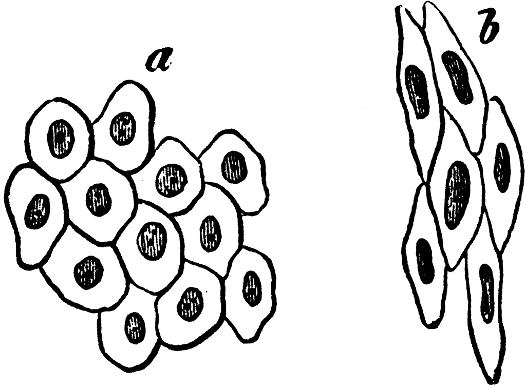
Simple Pavement Epithelium Cells | ClipArt ETC

White Blood Cells Diagram
7 Best Images of Neuron Label Worksheet - Blank Neuron Cell Diagram ...
Post a Comment for "39 human cell diagram with labels"