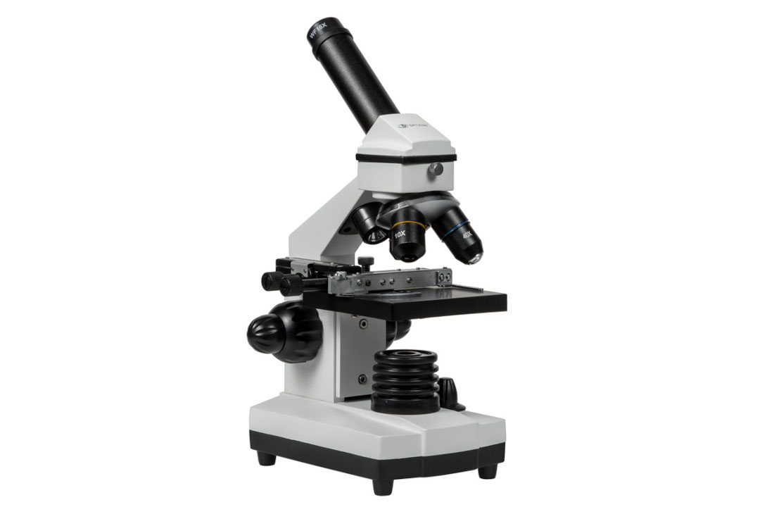
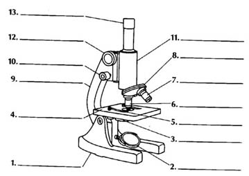
43 compound microscope unlabeled
microbenotes.com › flow-cytometryFlow Cytometry-Definition, Principle, Parts, Steps, Types, Uses The resulting cells are then incubated in test tubes or microtiter plates with unlabeled or fluorescently conjugated antibodies and analyzed through the flow cytometer machine. Antibody Staining. Once the sample is prepared, the cells are coated with fluorochrome-conjugated antibodies specific for the surface markers present on different cells. Joints Anatomy And Physiology Laboratory Manual Answers joints-anatomy-and-physiology-laboratory-manual-answers 3/21 Downloaded from ukc-mail.deskpro.com on August 14, 2022 by guest presentations, and more to help
Manganese Schiff Base Complexes, Crystallographic Studies, Anticancer ... The effect of the Mn(II) compound on the cellular viability was determined using the colorimetric MTT assay. The cells (1 × 10 5 cells/well) were cultured in a CO 2 incubator with 25 cm 3 tissue culture flasks from Nunc, Denmark, observed consistently under an inverted microscope for any contamination, and subsequently seeded into a 96-well ...

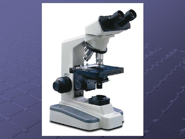
Compound microscope unlabeled
Schaftoside inhibits 3CLpro and PLpro of SARS-CoV-2 ... - ScienceDirect The compounds were injected as analytes at various concentrations at a flow rate of 30 μL/min with a contact time of 60 s and a dissociation time of 60 s, using PBS containing 5% DMSO and 0.05% Surfactant P20 as running buffer. Data were analyzed by the Biacore evaluation software. › protocols › immunofluorescenceImmunofluorescence Protocol Unlike direct immunofluorescence, indirect immunofluoresence is a double-layer technique. The unlabeled antibody is applied directly to the tissue substrate and then treated with a fluorochrome-conjugated anti-IgG. There are several advantages to this technique, and it is typically used more frequently than the direct method. Defining the ultrastructure of the hematopoietic stem cell niche by ... Zeiss LSM 880 confocal microscope was used to image the KM. ... Larvae were then flat embedded between glass slides coated with mould-release compound and left in an oven at 60°C for 72 hr. Larvae ... The identification of two unlabeled putative HSPCs in the same anatomical location further confirmed that HSPCs lodge in the perivascular KM ...
Compound microscope unlabeled. limitbauereddie 🙈That You Can Build Today You will be able to use this for years, because it is built to last.|Once opened, it extends to 55.5 inches, is 34.5 inches wide, and reaches to 24 inches tall. Perfect size for anyone. The seat ranges from 12.5 inches all the way up to 17 inches. An offset is included that ranges from 6.75 inches to 8.75 inches. Frontiers | 131I-Caerin 1.1 and 131I-Caerin 1.9 for the treatment of ... In this study, we used chloramine-T to couple 131 I with two high-affinity peptides (Caerin 1.1 and Caerin 1.9) and then analyzed the in vivo and in vitro effect of unlabeled Caerin 1.1, unlabeled Caerin 1.9, 131 I-labeled Caerin 1.1, and 131 I-labeled Caerin 1.9 for the treatment of NSCLC. Materials and methods Cell line and cell culture › books › NBK26880Looking at the Structure of Cells in the Microscope ... This latter quantity is a measure of the width of the entry pupil of the microscope, scaled according to its distance from the object; the wider the microscope opens its eye, so to speak, the more sharply it can see . Under the best conditions, with violet light (wavelength = 0.4 μm) and a numerical aperture of 1.4, a limit of resolution of ... Single-cell RNA-sequencing identifies anti-cancer immune phenotypes in ... Microenvironmental changes in the early metastatic niche may be exploited to identify therapeutic targets to inhibit secondary tumor formation and improve disease outcomes. We dissected the developing lung metastatic niche in a model of metastatic, triple-negative breast cancer using single-cell RNA-sequencing. Lungs were extracted from mice at 7-, 14-, or 21 days after tumor inoculation ...
Frontiers | Effects of Graded Whey Supplementation During Extreme ... We examined hypertrophic outcomes of weekly graded whey protein dosing (GWP) vs. whey protein (WP) or maltodextrin (MALTO) dosed once daily during 6 weeks of high-volume resistance training (RT). College-aged resistance-trained males (training age = 5 ± 3 years; mean ± SD) performed 6 weeks of RT wherein frequency was 3 d/week and each session involved 2 upper- and 2 lower-body exercises (10 ... Basic Microscope Diagram - microscope diagram purposegames, images 01 ... Basic Microscope Diagram - 15 images - label the neuron clip art at vector clip art online, microscope diagram fill online printable fillable blank pdffiller, animal anatomy biology4isc, images 01 introduction and terminology basic human anatomy, Flow Cytometry Is A Vital Step In The Sorting, Counting, And ... Following the staining step, cells are placed in tubes or microtiter plates and incubated with unlabeled or fluorescently-labeled antibodies before being analysed on a flow cytometer. A laser beam is used in Flow Cytometry, an analytical instrument, to measure and examine a number of physical properties of cells or particles dispersed in a fluid. De novo pyrimidine synthesis is a targetable vulnerability in IDH ... We recently created isogenic IDH1-mutant and IDH1 wild-type (WT) glioma cell culture models that recapitulate (R)-2HG levels in primary brain tumors (McBrayer et al., 2018).Using an endogenous IDH1/2 WT human glioma line, HOG, we expressed the IDH1-R132H oncogene (HOG-R132H) or an empty vector (HOG-EV). We used these isogenic models with a compound screening platform developed by two of us (J ...
microbeonline.com › indirect-fluorescent-antibodyIndirect Fluorescent Antibody (IFA) Test • Microbe Online May 21, 2022 · Again the excess antibody is removed by washing and the smear is visualized using a fluorescence microscope. DFA and IFA for the detection of viral antigen (Image source:virology.ws) Comparison between IFA and DFA. IFA is more sensitive than Direct Fluorescent Assay (DFA) because the second antibody is coupled to the indicator. Bio 4150 Human Anatomy and Physiology Course Procedure Label the segments of an unlabeled ECG and match each event to a cardiac cycle occurrence. Given any two of three variables (cardiac output, heart rate, and stroke volume), solve for the value of the third. Given a random list of cardiac cycle events, rank the list in their logical order of occurrence. Explain the functions of the lymphatics. Antioxidants | Free Full-Text | Bacterial Metabolite Reuterin ... Reuterin is well-known for its broad-spectrum antimicrobial ability, while the other potential bioactivity is not yet clear. The present study aims to investigate the immunomodulatory activity of reuterin on chicken macrophage HD11 cells for the first time and evaluate whether reuterin is able to regulate the lipopolysaccharide-stimulated inflammatory response. The results showed that the safe ... how can a light microscope work - Be Refined Site Gallery Of Photos We offer the best alternatives to conventional halogen cold light sources for microscopes. A simple light microscope manipulates how light enters the eye using a convex lens where both sides of the lens are curved outwards. Magnification can be achieved in a microscope by moving one or more lenses to different focal lengths.
Label the microscope — Science Learning Hub
La Semplice enigmistica per bambini & bambine La Semplice enigmistica per bambini & bambine Latest Journal's Impact IF 2021-2022| Trend, Prediction, Ranking & Key Factor Analysis - Academic Accelerator

Sci10U3L2
Timing of neuron development determines what they can become To do this, the researchers labeled the precursor cells at different times so that all neurons that emerged from these precursors in the following days lit up green under the microscope. In...

Microscope Diagram Labeled, Unlabeled and Blank | Parts of a ...
› protocol-and-troubleshooting › iimmunohistochemistry Principle. How IHC staining Works View the coverslip under fluorescence microscope; Tips: Operations of Fluorescence Microscope. Operate the microscope according to the manual; Turn on the mercury lamp for 5-15 min to stabilize the light source before use; Wear protective glasses when adjusting light source to avoid harmful ultraviolet rays to eyes

Optics Optical Microscope, Rs 4500/piece SD Optical Lab India ...
Cryo-EM structures of Escherichia coli Ec86 retron complexes reveal ... Here, we investigate the Escherichia coli Ec86 retron and use cryo-electron microscopy to determine the structures of the Ec86 (3.1 Å) and cognate effector-bound Ec86 (2.5 Å) complexes. The Ec86...

Microscopy | biology
microscope | Types, Parts, History, Diagram, & Facts Optical microscopes can be simple, consisting of a single lens, or compound, consisting of several optical components in line. The hand magnifying glass can magnify about 3 to 20×. Single-lensed simple microscopes can magnify up to 300×—and are capable of revealing bacteria —while compound microscopes can magnify up to 2,000×.

Microscope Diagram Labeled, Unlabeled and Blank | Parts of a ...
Olympus' new generation intelligent digital imaging system APX100 ... GC allows users to obtain three-dimensional light and shadow effects when observing unlabeled samples. Compared with phase contrast (PH), GC images have no halo phenomenon that affects accurate observation; unlike differential interference (DIC), which only allows observation through glass vessels .
Opticon Biolife 1024x microscope - white Botland - Robotic Shop
Antagonism of the Muscarinic Acetylcholine Type 1 Receptor Enhances ... Expression of M 1 R in Human Neuroblastoma SH-SY5Y Cell Line and Rodent DRG Neurons. The presence of M 1 R in SH-SY5Y cell line and rat DRG neurons was assessed by using MT7-ATTO590 (Fig. 1A-D).This labelled MT7 is absolutely specific for the M 1 R and is superior to the use of antibodies that cross-react with other MR sub-types. The specificity of MT7-ATTO590 was confirmed by using DRG ...

ScienceSupply.com (@Science_Supply) / Twitter
A conformational change controlling the toxicity of the prion protein ... Adeno-associated virus manufacturing and in vivo transductionSingle-stranded adeno-associated virus (ssAAV) vector backbones with AAV2 inverted terminal repeats (ITRs) had been kindly supplied by B. Schneider (EPFL). Herein, expression of the monomeric NeonGreen fluorophore was pushed by the human Synapsin I (hSynI) promoter. A P2A sequence (GSGATNFSLLKQAGDVEENPGP) was launched between mNG and ...
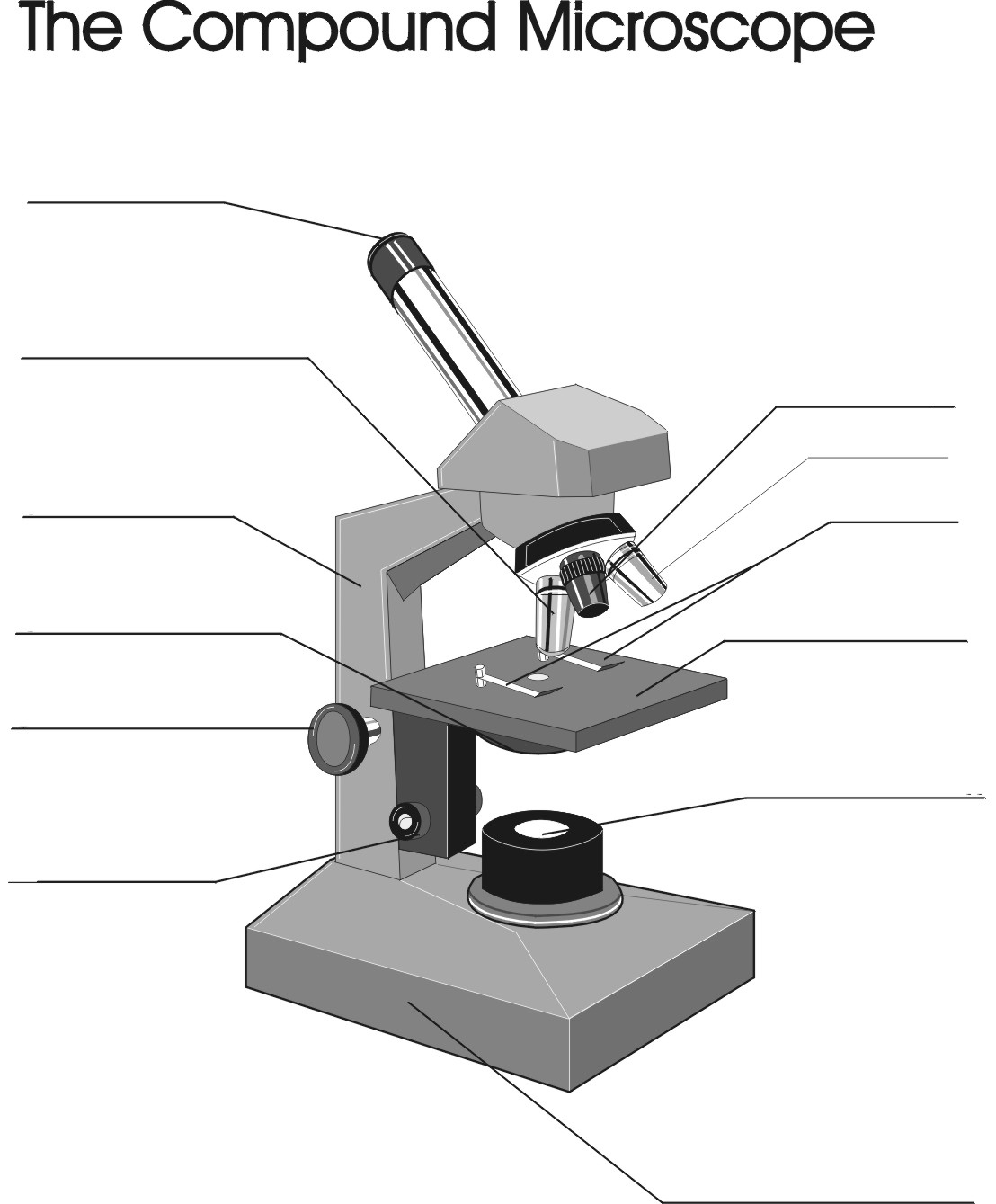
Copy of "The Compound Microscope"
› science › articleCopper-mediated radioiodination and radiobromination via aryl ... Using a copper-mediated radioiodination method, we synthesized [125 I]mIB-PS via the boronic precursor (3) in method I (Scheme 2A).The reaction was conducted under conditions described in our previous report, with some modifications. 12 A boronic precursor 3, Cu(py) 4 (OTf) 2 (py = pyridine, OTf = trifluoromethanesulfonate), and [125 I]NaI aqueous solution were added to a microtube.
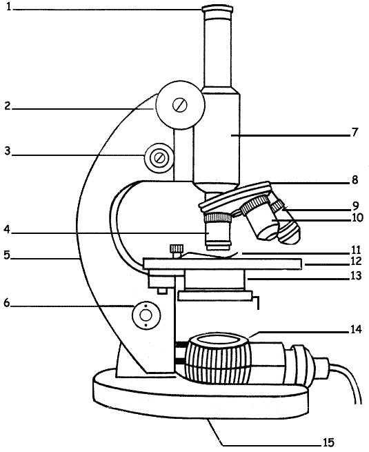
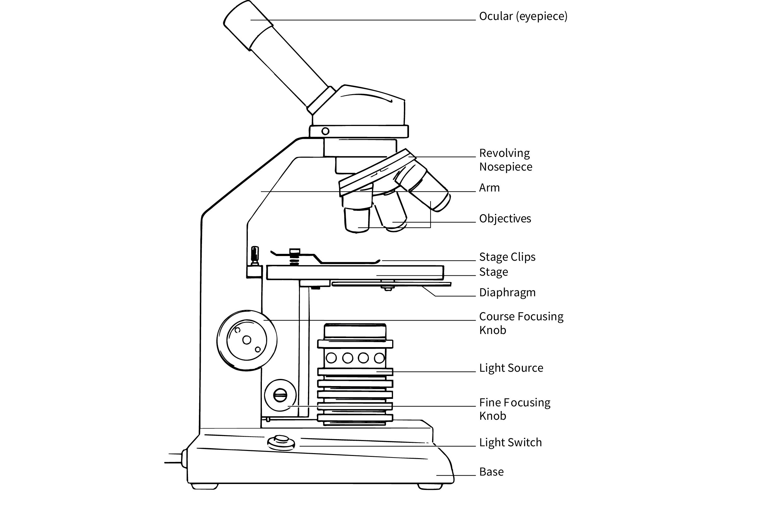
The Microscope Parts and Use
Impaired disassembly of the axon initial segment restricts ... Notably, this result was not due to unlabeled mitochondria in Rpt3 CKO mice because the results shown in Fig ... The images are acquired with lightsheet 7 microscope (Zeiss, Germany). For CUBIC treated brain and spinal cord, images were captured using a 5 × objective lens with digital zoom from 0.8 × to 2.5 × with digital zoom equipped with ...

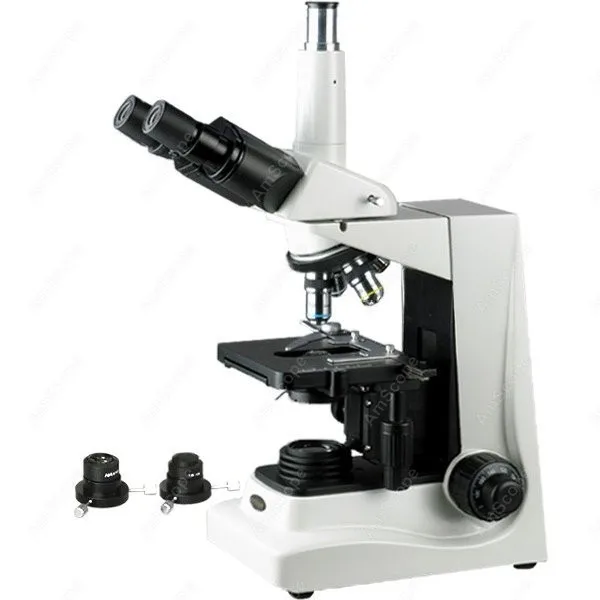
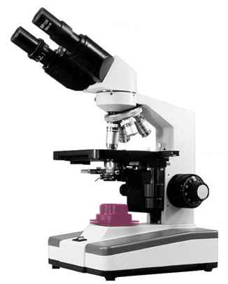
Compound Microscope
Non-canonical function of an Hif-1α splice variant contributes to the ... Unlabeled probes were used as competitors. SH represents shift binding of Hif-1α2, UB represents unspecific banding. ... (RT) PBS. The washed tissue was then embedded in optimal cutting temperature compound and immediately placed on dry ice and sectioned at -20°C. Subsequently, 15 µm sections were mounted on microscope glass slides (Thermo ...

Compound Microscope Parts – Labeled Diagram and their ...
Identifying cell receptors for the nanoparticle protein corona using ... Images were acquired using a Leica SP8 Confocal microscope at The Imaging Facility at The Hospital for Sick Children. STRING The nanoparticle-adsorbed proteins and LDLR were input into the STRING ...

40X-1000X Binocular Compound Laboratory Microscope with Blank ...
Pharmaceutics | Free Full-Text | Development and Evaluation of a ... In addition, the uptake of 68 Ga-yG5-RGD in BxPC3 was significantly blocked by excess amounts of AMD3100 (an FDA-approved CXCR4 antagonist) and/or unlabeled RGD ( p < 0.001), confirming its dual-receptor targeting properties. The ex vivo biodistribution and immunohistochemical results were consistent with the in vivo imaging results.

Microscope Diagram Labeled, Unlabeled and Blank | Parts of a ...
› en › microscopeFundamental Concepts in DIC Microscopy - Life Science The relationship between optical path gradients and intensity profiles in DIC microscopy is illustrated in Figure 2. The specimen presented in Figure 2(a) is a doughnut-shaped human erythrocyte imaged at high magnification in differential interference contrast with the shear axis indicated by a double-headed arrow (northwest to southeast).
Darkfield Brightfield Trinocular Microscope--AmScope Darkfield Brightfield Trinocular Compound Microscope 40X-1600X T600A-DK
backyardgrillsmoker 😪Projects That Sell Well backyardgrillsmoker 😋Round Yard. Overall, get creative! There are so many unique DIY planter ideas out there and so many really adorable ones (like these terrarium planters!) available to tune your indoor apartment garden to your taste.''}'''''Shank 3.175mm TOY JEWELRY MAKING, Crafts, CNC, PCB, Woodworking, MODEL TRAINS CARS BOATS!! 4.7 out of 5 stars 7 $29.95 End Mill Sets, Tungsten Steel ...

Mikroskop optik Leica Microsystems Digital microscope Fase ...
24 questions with answers in BACTERIAL RESISTANCE - ResearchGate When we say that one of the reasons for having bacterial resistance is because of the use of antibiotics in cattle, so I wonder if: 1. This issue is related to resistant bacteria selection in the ...
Microscope Challenge - Andrew Herrick: Teaching Portfolio
Clumps of mesenchymal stem cells/extracellular matrix complexes ... The sections were observed using the NIKON ECLIPSE E600 microscope (NIKON, Tokyo, Japan). To detect the PKH26-labeled cells and rat COL1, an immunofluorescence analysis was conducted. Briefly, the fixed specimens were embedded in Tissue-Tek OCT compound, and 20-μm-thick serial sections were cut using a cryostat.
COMPOUND LIGHT MICROSCOPE USES LIGHT PASSING THROUGH THE
Defining the ultrastructure of the hematopoietic stem cell niche by ... Zeiss LSM 880 confocal microscope was used to image the KM. ... Larvae were then flat embedded between glass slides coated with mould-release compound and left in an oven at 60°C for 72 hr. Larvae ... The identification of two unlabeled putative HSPCs in the same anatomical location further confirmed that HSPCs lodge in the perivascular KM ...
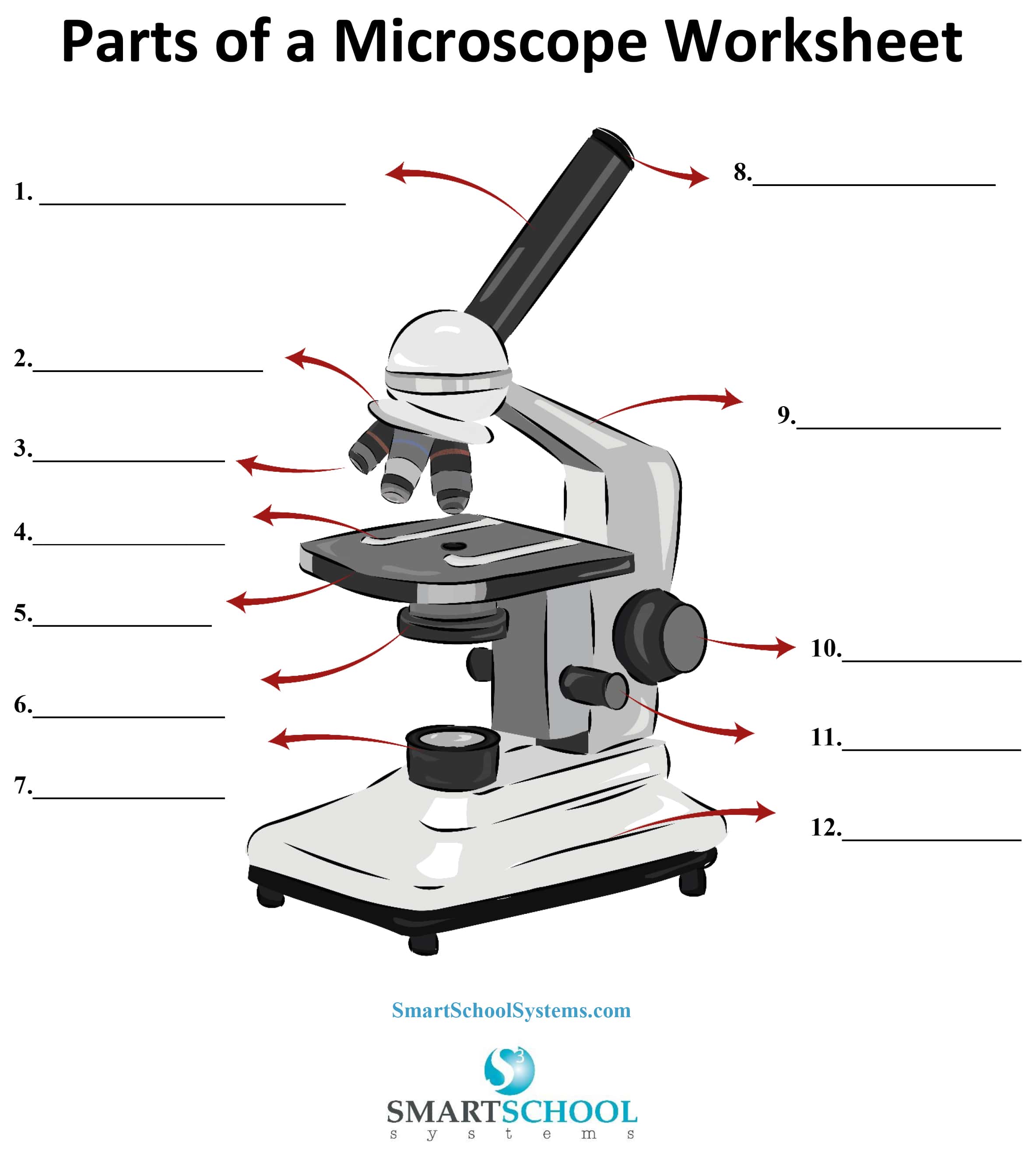
Parts of a Microscope - SmartSchool Systems
› protocols › immunofluorescenceImmunofluorescence Protocol Unlike direct immunofluorescence, indirect immunofluoresence is a double-layer technique. The unlabeled antibody is applied directly to the tissue substrate and then treated with a fluorochrome-conjugated anti-IgG. There are several advantages to this technique, and it is typically used more frequently than the direct method.
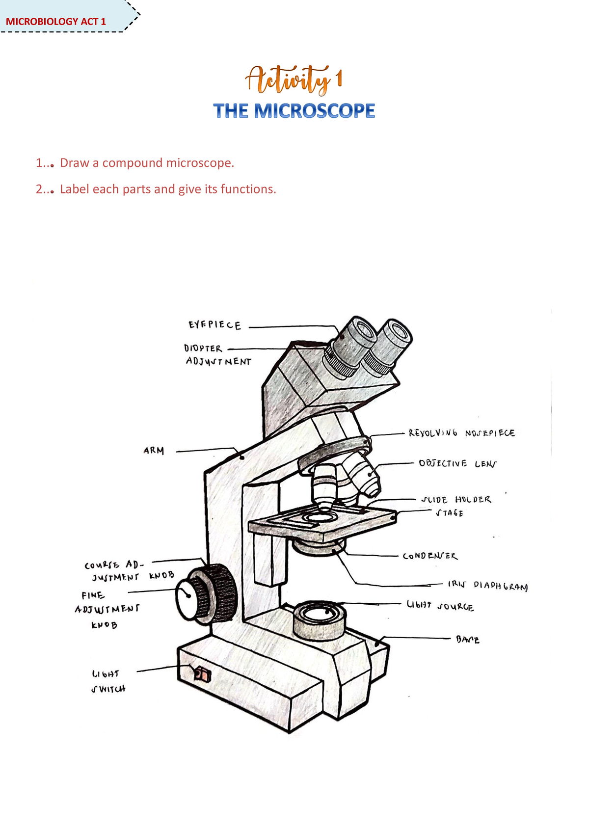
Microscope Activity - MICROBIOLOGY - 1... Draw a compound ...
Schaftoside inhibits 3CLpro and PLpro of SARS-CoV-2 ... - ScienceDirect The compounds were injected as analytes at various concentrations at a flow rate of 30 μL/min with a contact time of 60 s and a dissociation time of 60 s, using PBS containing 5% DMSO and 0.05% Surfactant P20 as running buffer. Data were analyzed by the Biacore evaluation software.

Transmitted light microscope OBE-12 • 13 - KERN & SOHN GmbH

File:Olympus CH2 microscope 1.jpg - Wikimedia Commons

Microscope Use Lab Purpose: To learn the parts and how to use ...

Introduction to the Compound Light Microscope Chuck Hesbacker ...
blank diagram of a compound light microscope - Clip Art Library
Microscope Parts Labeled - ClipArt Best
What is a Compound Microscope? | Flinn Scientific

Compound Microscope Diagram Game

Leica DM100 Microscope | Monocular Student Microscope ...

OMAX 2500X LED Binocular Compound Microscope+Blank Slides+Covers+Lens Paper+Book

microscope clipart - Clip Art Library

Label Microscope Diagram - EnchantedLearning.com

The Compound Microscope parts & how ... | Microscope parts ...
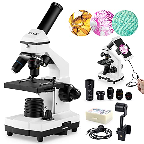
Best Compound Microscopes - Buying Guide | Gistgear

Microscope Labeling

40X-400X Monocular Student Biological Microscope for Beginners (BM-42)

Microscope With Labels Clip Art at Clker.com - vector clip ...

$500 to $1,000 – Tagged "Brand_National" – Microscope Central

Amazon.com: AmScope 40X-2000X LED Monocular Digital Compound ...

Microscope Components - Science Quiz

Optical microscope - MSC-B103T(Siedentopf) - Bioevopeak ...

Quotes about Microscope (115 quotes)
Using Microscopes
Post a Comment for "43 compound microscope unlabeled"