45 spirogyra microscope labeled
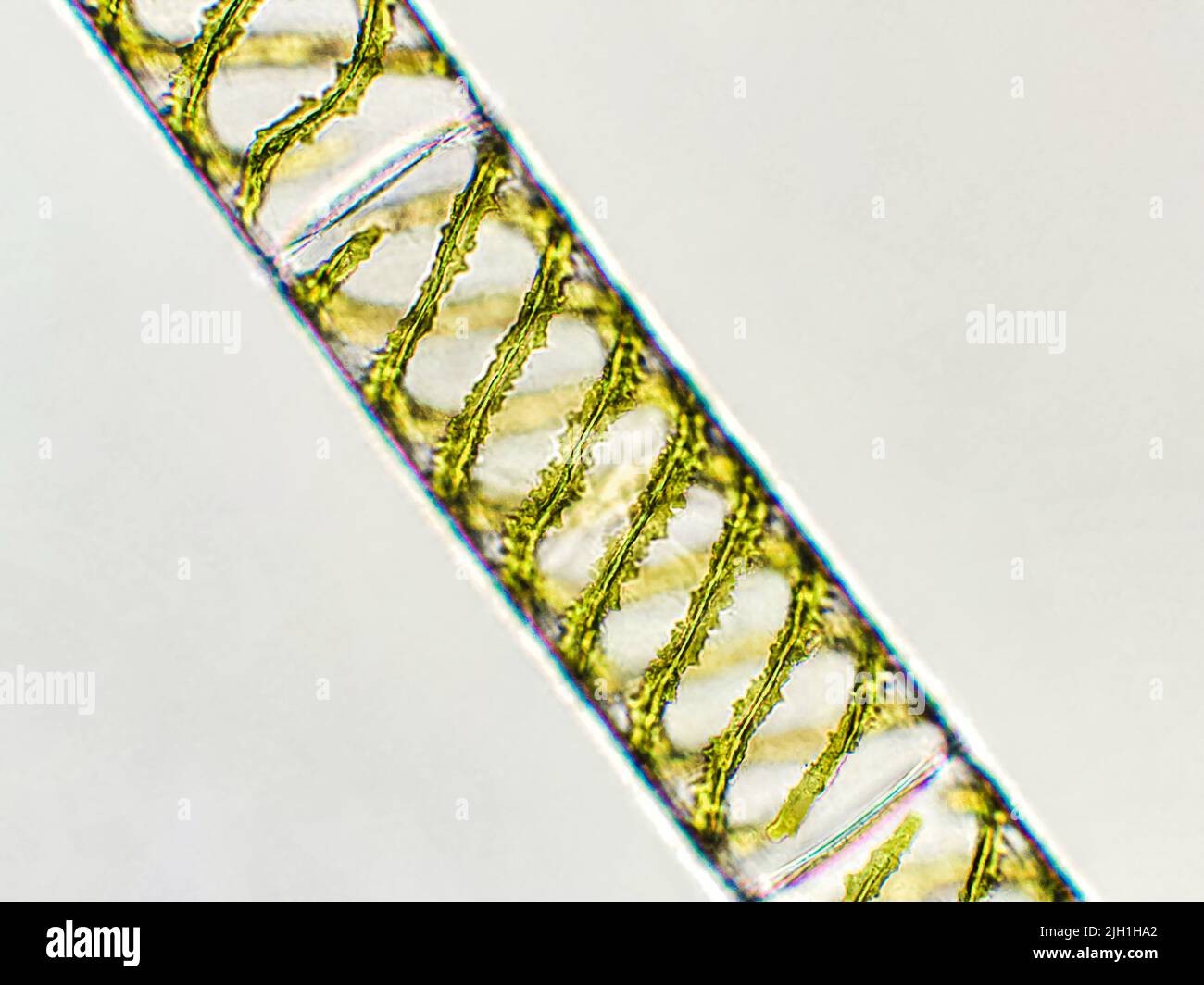
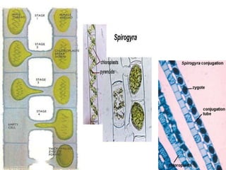
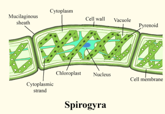
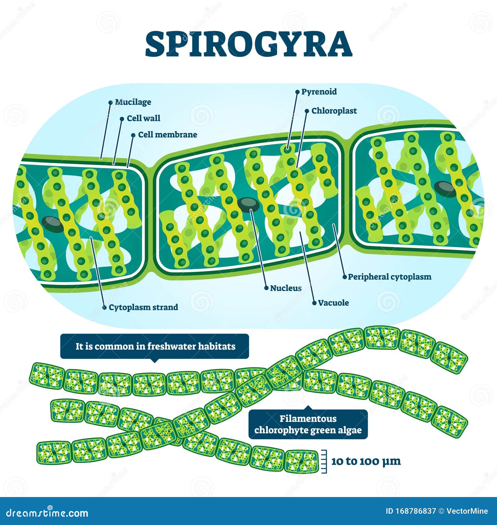
Spirogyra: Structure & Characteristics with Labeled Diagram What is spirogyra and how the cell looks like under a microscope: learn its characteristics - size & shape, reproduction, & lifecycle using facts & labeled ... Spirogyra algae | Galleries | Nikon Europe B.V. A1 HD25 / A1R HD25 - Spirogyra algae · Spirogyra algae · Produits de microscope Nikon · Solutions · Service après vente & Soutien · Liens · Autres Produits Nikon.
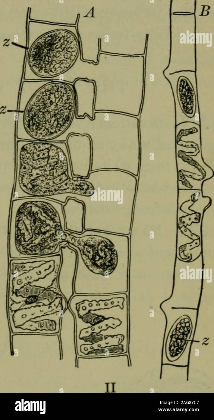
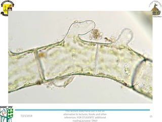
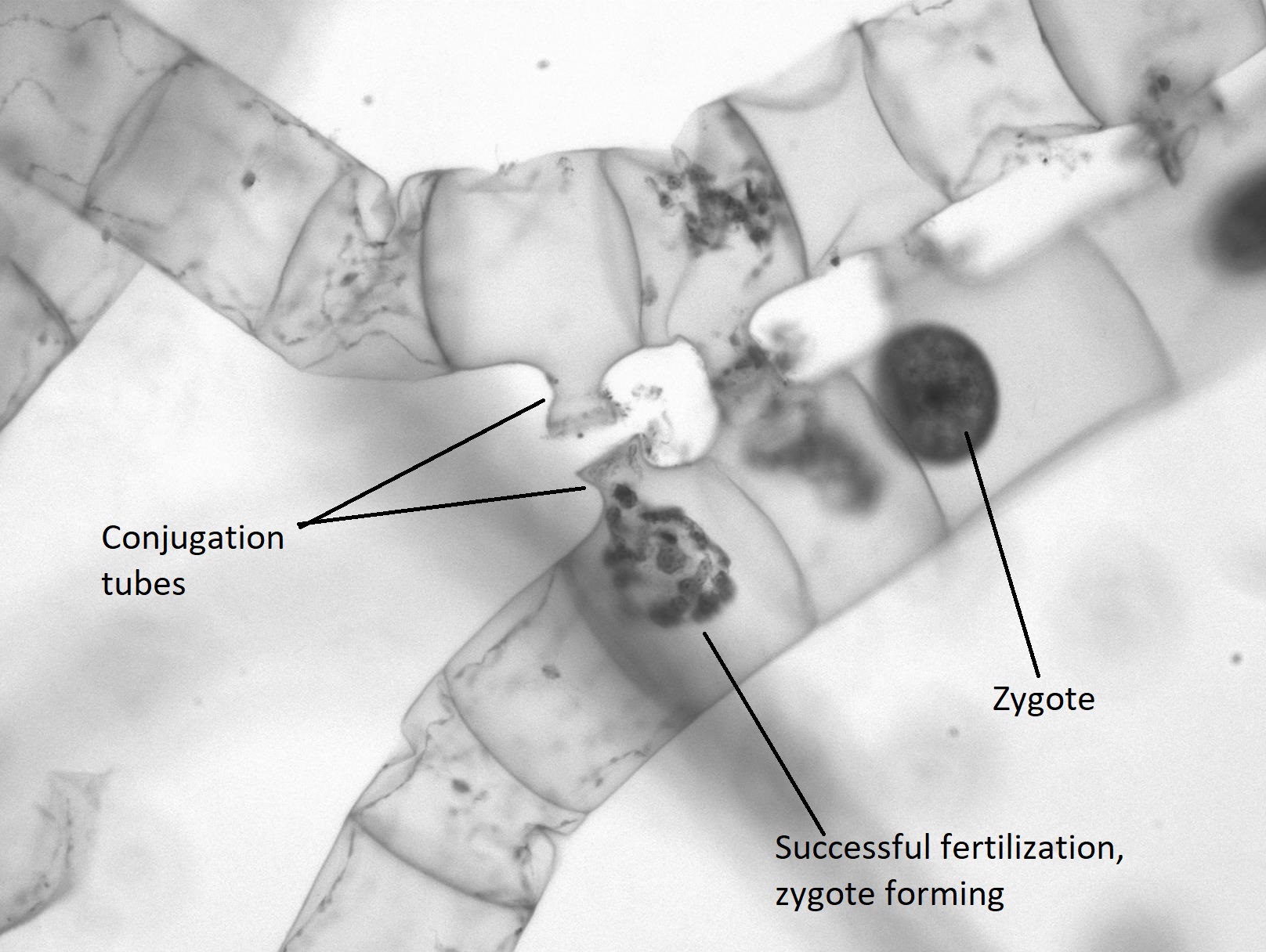
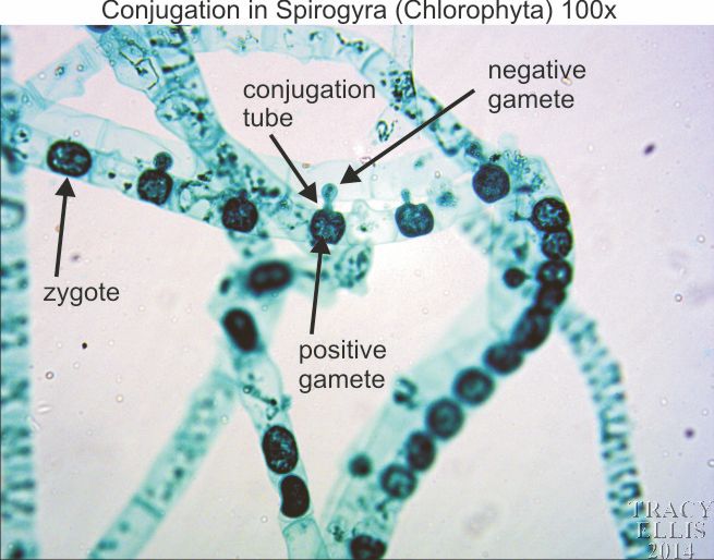
Spirogyra conjugation 100x - Dissection Connection Labelled microscope slide images prepared by Tracy John Ellis. These images are hosted only and no technical support will be available.
Spirogyra microscope labeled
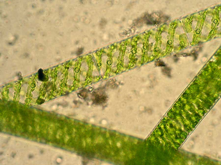
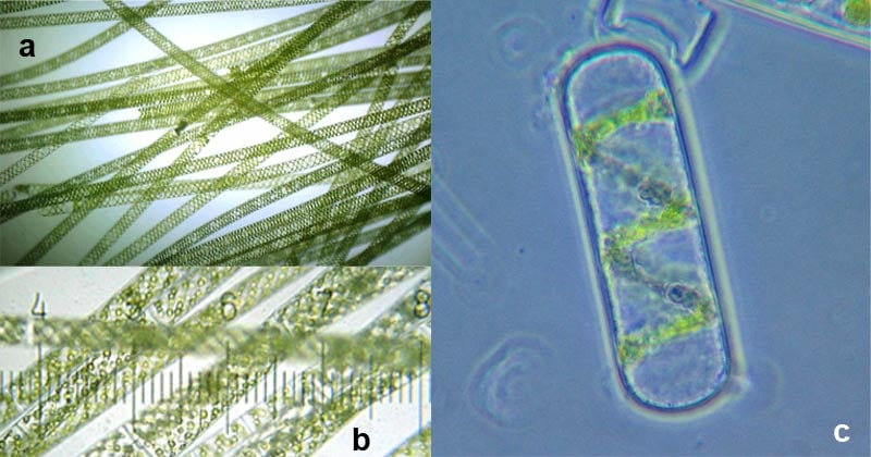
File:Spirogyra Under Light Microscope.jpg - Wikimedia Commons Nov 24, 2017 ... English: A filamentous green algae. Known for it's helical chloroplast structure. Date, 18 October 2017. Source, Own work. Author ... Spirogyra under the Microscope Jan 22, 2014 ... Spirogyra green algae captured under the biological microscope at both 100x and 400x magnification. Info and images. Draw a neat diagram of spirogyra and label on it - Vedantu In a microscopic view we can see the various parts of the cell of spirogyra. Cell wall is the outermost layer of a plant cell. Pyrenoid is a structure inside or ...
Spirogyra microscope labeled. Labeled Diagram of Spirogyra - Pinterest Nov 26, 2017 - Spirogyra is a sophisticated, filamentous green alga, ... good thing I have this electron microscope laying around | See more about Barbados, ... Spirogyra Under The Microscope - YouTube Aug 24, 2017 ... Spirogyra is a filamentous green algae found in freshwater environments. It is often found as green clumps, although each strand is microscopic. Spirogyra - Under the Microscope - YouTube Aug 24, 2016 ... Music: C Major Prelude - Bach. The following diagram illustrates a filament of spirogyra as seen ... Jan 25, 2020 ... The following diagram illustrates a filament of spirogyra as seen under the microscope. Its parts have been labelled as A,B,C,D. Functions ...
Draw a neat diagram of spirogyra and label on it - Vedantu In a microscopic view we can see the various parts of the cell of spirogyra. Cell wall is the outermost layer of a plant cell. Pyrenoid is a structure inside or ... Spirogyra under the Microscope Jan 22, 2014 ... Spirogyra green algae captured under the biological microscope at both 100x and 400x magnification. Info and images. File:Spirogyra Under Light Microscope.jpg - Wikimedia Commons Nov 24, 2017 ... English: A filamentous green algae. Known for it's helical chloroplast structure. Date, 18 October 2017. Source, Own work. Author ...













Post a Comment for "45 spirogyra microscope labeled"