39 label parts of microscope
What is a Compound Microscope? | Microscope World Blog The Parts & Function of a Compound Microscope A compound microscope is a high power (high magnification) microscope that uses a compound lens system. A compound microscope has multiple lenses: the objective lens (typically 4x, 10x, 40x or 100x) is compounded (multiplied) by the eyepiece lens (typically 10x) to obtain a high magnification of 40x, 100x, 400x and 1000x. PDF Parts of a Microscope Printables - Homeschool Creations Parts of a eyepiece arm stageclips nosepiece focusing knobs illuminator stage objective lenses head base Label the parts of the microscope. You can use the word bank below to fill in the blanks or cut and paste the words at the bottom. Microscope Created by Jolanthe @ HomeschoolCreations.net eyepiece head objective lenses arm focusing knob base ...
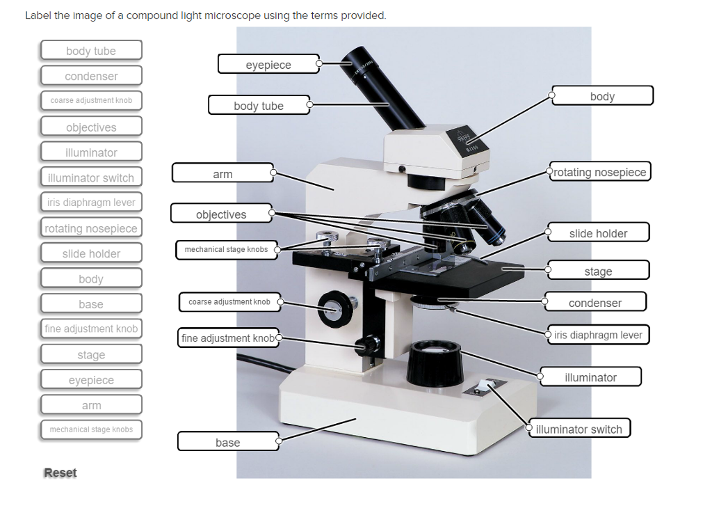
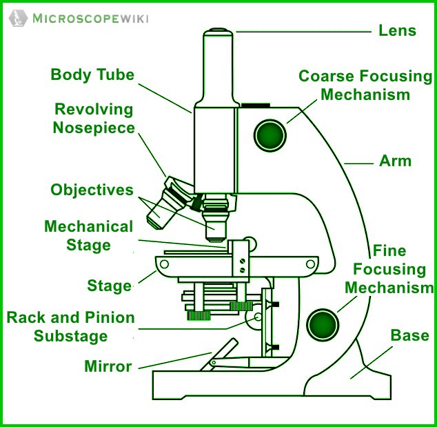
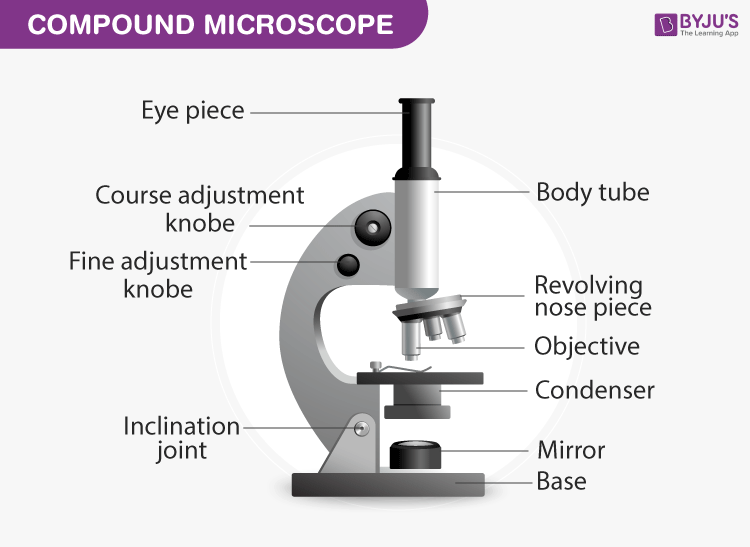
Compound Microscope Parts, Functions, and Labeled Diagram The individual parts of a compound microscope can vary heavily depending on the configuration & applications that the scope is being used for. Common compound microscope parts include: Compound Microscope Definitions for Labels Eyepiece (ocular lens) with or without Pointer: The part that is looked through at the top of the compound microscope ...

Label parts of microscope
Labeling the Parts of the Microscope | Microscope activity, Science ... Jan 13, 2016 - Free worksheets for labeling parts of the microscope including a worksheet that is blank and one with answers. Microscope Parts, Function, & Labeled Diagram - slidingmotion Microscope Parts Labeled Diagram The principle of the Microscope gives you an exact reason to use it. It works on the 3 principles. Magnification Resolving Power Numerical Aperture. Parts of Microscope Head Base Arm Eyepiece Lens Eyepiece Tube Objective Lenses Nose Piece Adjustment Knobs Stage Aperture Microscopic Illuminator Condenser Lens Microscope, Microscope Parts, Labeled Diagram, and Functions Jan 19, 2022 · Microscope cell staining is a technique used to improve the visibility of cells and cell parts under a microscope. A nucleus or a cell wall can be seen more clearly by using different stains. 2. Iodine, crystal violet, and methylene blue are examples of simple stains. 3. Make a wet or dry mount with a coverslip. 4.
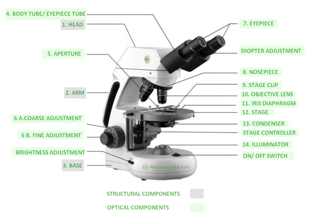
Label parts of microscope. Binocular Microscope Anatomy - Parts and Functions with a Labeled ... Optical parts of the microscope The microscope's condenser is the structure that collects and focuses light from the light source. You will see an aperture in the condenser that controls the amount of light coming through it. A standard light microscope possesses three to five objective lenses that range in power from 10X to 100X. Parts of the Microscope with Labeling (also Free Printouts) Table of Contents 1. Eyepiece 2. Body tube/Head 3. Turret/Nose piece 4. Objective lenses 5. Knobs (fine and coarse) 6. Stage and stage clips 7. Aperture 9. Condenser 10. Condenser focus knob 11. Iris diaphragm 12. Diopter adjustment 13. Arm 14. Specimen/slide 15. Stage control/stage height adjustment 16. On and off switch 17. Base Label the microscope — Science Learning Hub 08.06.2018 · All microscopes share features in common. In this interactive, you can label the different parts of a microscope. Use this with the Microscope parts activity to help students identify and label the main parts of a microscope and then describe their functions.. Drag and drop the text labels onto the microscope diagram. If you want to redo an answer, click on the … Parts Of A Microscope Labeling Teaching Resources | TpT This resource contains 1 worksheet for students to label the parts of a microscope. Answer key included. File comes as both a PDF for printing and a Google Slides resource for a digital classroom where students can type in answers if you wish. This resource can be used as an introduction to new material or a study guide for a quiz.
Microscopy Pre-lab Activities - University of Delaware Microscope controls: turn knobs (click and hold on upper or lower portion of knob) throw switches (click and drag) turn dials (click and drag) move levers (click and drag) changes lenses (click and drag on objective housing) select a specimen (click on a slide) adjust oculars (in "through" view, with the light on, click and drag to move oculars closer or further apart) … Microscope Labeling Game - PurposeGames.com About this Quiz. This is an online quiz called Microscope Labeling Game. There is a printable worksheet available for download here so you can take the quiz with pen and paper. This quiz has tags. Click on the tags below to find other quizzes on the same subject. Science. Label The Parts Of A Microscope Teaching Resources | TpT Given a collection of 25 total draggable items, students must place them in their correct location on the microscope by a "drag and drop" modeling activity. Students are provided 12 microscope parts and 13 labels of the parts. Upon placing the parts in their accurate location, students must drag the labels next to their correct components. Compound Microscope Parts - Labeled Diagram and their Functions There are three major structural parts of a compound microscope. The head includes the upper part of the microscope, which houses the most critical optical components, and the eyepiece tube of the microscope. The base acts as the foundation of microscopes and houses the illuminator. The arm connects between the base and the head parts.
Parts of Stereo Microscope (Dissecting microscope) – labeled … Optical parts of a stereo microscope work together to magnify and produce a 3-D image of the specimens. These parts include: Eyepieces. The eyepiece (or ocular lens) is the lens part at the top of a microscope that the viewer looks through. Typically, standard eyepieces for a dissecting microscope have a magnifying power of 10x. Optional eyepieces of varying powers are … PDF Label parts of the Microscope: Answers Label parts of the Microscope: Answers Coarse Focus Fine Focus Eyepiece Arm Rack Stop Stage Clip . Created Date: 20150715115425Z ... PDF Label parts of the Microscope Label parts of the Microscope: . Created Date: 20150715115425Z label parts of a microscope Quiz - PurposeGames.com This is an online quiz called label parts of a microscope There is a printable worksheet available for download here so you can take the quiz with pen and paper. From the quiz author label the correct names of the parts of the microscope This quiz has tags. Click on the tags below to find other quizzes on the same subject. microscope microscopy
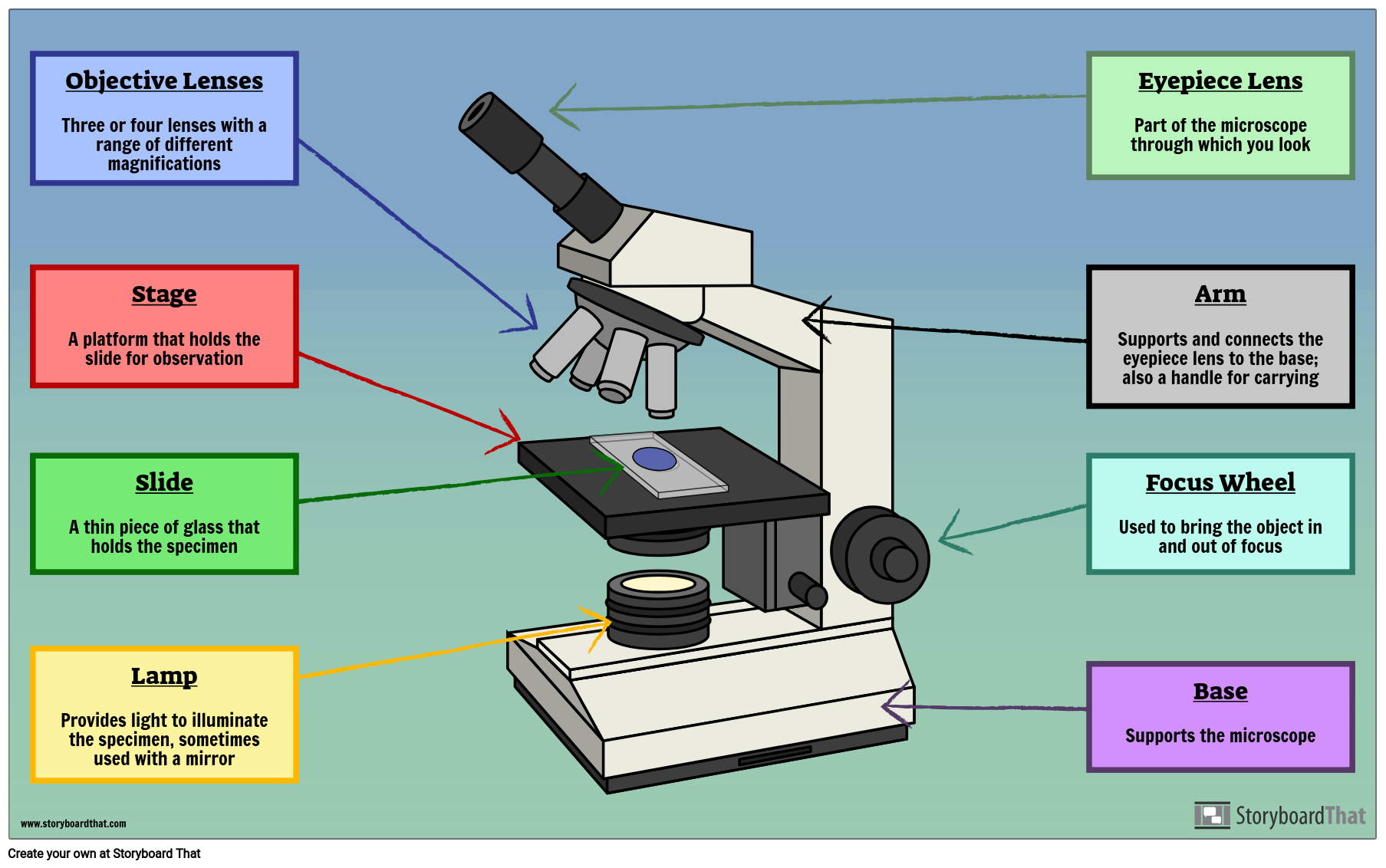
Microscope Parts & Functions - AmScope Objective Lenses: Usually you will find 3 or 4 objective lenses on a microscope. The most common ones are 4X (shortest lens), 10X, 40X and 100X (longest lens). The higher power objectives (starting from 40x) are spring loaded.
Fluorescence Microscopy - Explanation and Labelled Images 16.12.2020 · The main parts of a fluorescent microscope overlap with the traditional light microscope. However, there are two main features that sets fluorescent microscope apart from the traditional microscope. One is the type of light source and the other is the use of specialized filter elements. A fluorescence microscope uses a higher intensity light to illuminate the …
Parts of a Microscope - The Comprehensive Guide Microscope Parts Labeled: Parts of A Microscope 1. Eyepiece Lens and Eyepiece Tube 2. Objective Lens 3. Tube 4. Base 5. Arm 6. Illuminator 7. Stage or platform 8. Stage Clips 9. Rotating Turret or Nosepiece 10. Rack Stop 11. Condenser Lens 12. Iris or Diaphragm 13. Coarse Adjustment Knob 14. Fine Adjustment Knob 15. Power Switch
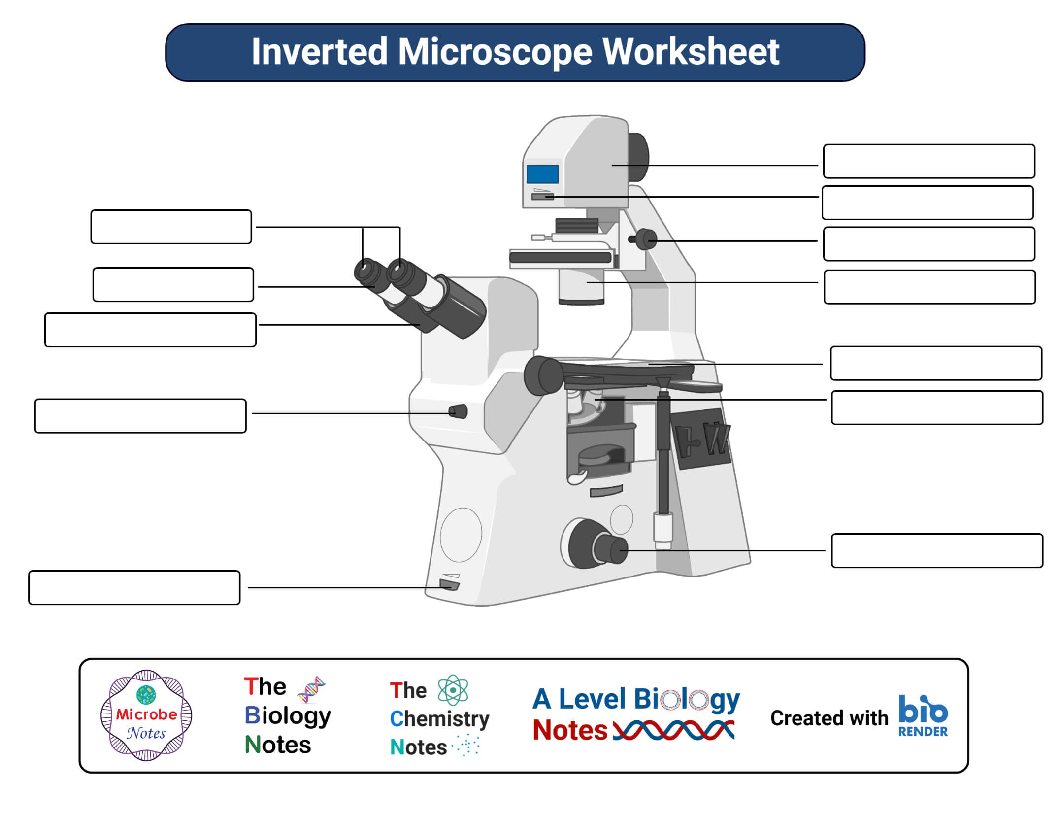
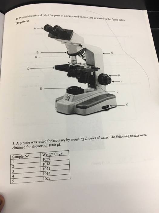
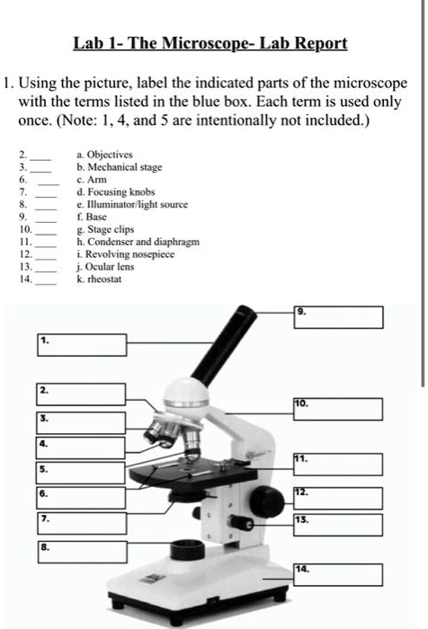
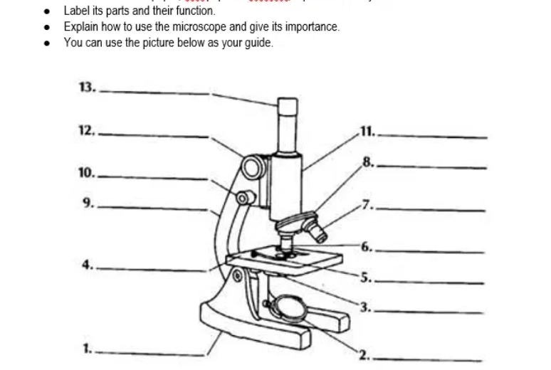
Labeling the Parts of the Microscope | Microscope World Resources Labeling the Parts of the Microscope This activity has been designed for use in homes and schools. Each microscope layout (both blank and the version with answers) are available as PDF downloads. You can view a more in-depth review of each part of the microscope here. Download the Label the Parts of the Microscope PDF printable version here.
Microscope Parts and Functions Most specimens are mounted on slides, flat rectangles of thin glass. The specimen is placed on the glass and a cover slip is placed over the specimen. This allows the slide to be easily inserted or removed from the microscope. It also allows the specimen to be labeled, transported, and stored without damage.
Virtual Microscope - NCBioNetwork.org Lesson Description BioNetwork’s Virtual Microscope is the first fully interactive 3D scope - it’s a great practice tool to prepare you for working in a science lab. Explore topics on usage, care, terminology and then interact with a fully functional, virtual microscope. When you are ready, challenge your knowledge in the testing section to see what you have learned.
UD Virtual Compound Microscope - University of Delaware ©University of Delaware. This work is licensed under a Creative Commons Attribution-NonCommercial-NoDerivs 2.5 License.Creative Commons Attribution-NonCommercial-NoDerivs 2.5 …
Antonie van Leeuwenhoek, Father of Microbiology - ThoughtCo 21.07.2019 · Known For: Improvements to the microscope, discovery of bacteria, discovery of sperm, descriptions of all manner of microscopic cell structures (plant and animal), yeasts, molds, and more; Also Known As: Antonie Van Leeuwenhoek, Antony Van Leeuwenhoek; Born: Oct. 24, 1632 in Delft, Holland; Died: Aug. 30, 1723 in in Delft, Holland; Education: Only basic education
Label parts on microscope Flashcards | Quizlet ashleibryson Terms in this set (14) Eyepiece Body Tube on microscope High power objective Low power objective Stage Opening Diaphragm Lever Light on microscope Coarse adjustment Fine adjustment Nosepiece Arm on a microscope Stage Clip Stage on microscope Base Sets found in the same folder Cell Organelles 28 terms MKL67 Parts of a Microscope
Parts of a microscope with functions and labeled diagram 19.04.2022 · Thank you very much it really helped me with my science home work since i in 8th grade and this my home work to draw a microscope label all the parts and the function thank may the holy father of holy spirits bless you and give more wisdom thanks love you all keep up the good work and thank you again bye. Reply . Sagar Aryal. May 21, 2022 at 1:57 AM . Thank you …
Microscope, Microscope Parts, Labeled Diagram, and Functions Jan 19, 2022 · Microscope cell staining is a technique used to improve the visibility of cells and cell parts under a microscope. A nucleus or a cell wall can be seen more clearly by using different stains. 2. Iodine, crystal violet, and methylene blue are examples of simple stains. 3. Make a wet or dry mount with a coverslip. 4.
Microscope Parts, Function, & Labeled Diagram - slidingmotion Microscope Parts Labeled Diagram The principle of the Microscope gives you an exact reason to use it. It works on the 3 principles. Magnification Resolving Power Numerical Aperture. Parts of Microscope Head Base Arm Eyepiece Lens Eyepiece Tube Objective Lenses Nose Piece Adjustment Knobs Stage Aperture Microscopic Illuminator Condenser Lens
Labeling the Parts of the Microscope | Microscope activity, Science ... Jan 13, 2016 - Free worksheets for labeling parts of the microscope including a worksheet that is blank and one with answers.














Post a Comment for "39 label parts of microscope"